Central Venous Pressure Waveform
A central line in the superior vena cava records central venous pressure (CVP), which approximates right atrial pressure. Its waveform has three positive waves and two descents, each tied to an event in the cardiac cycle, and the shape changes in recognizable ways with arrhythmias, valve disease, and pericardial disease. This page labels each component against the ECG and shows the common abnormal patterns.
Last reviewed October 8, 2026. A study and documentation reference.
On this page: the waves · measuring CVP · abnormal patterns · central line basics · what CVP cannot tell you · in a note
The waves and descents
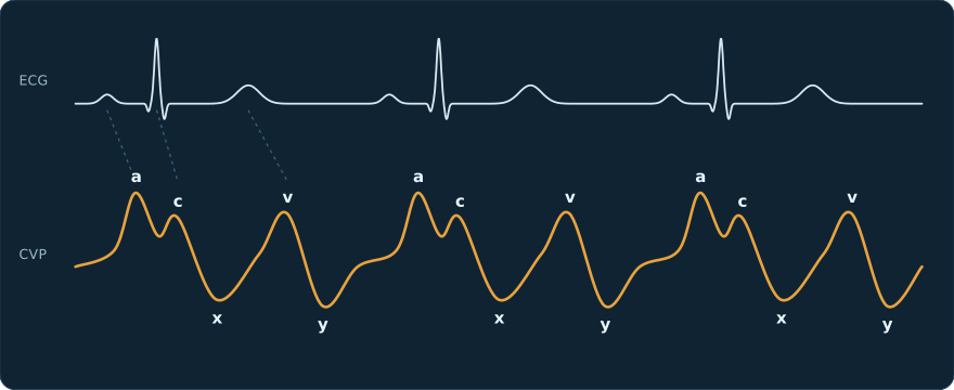
| Component | Cardiac event | ECG timing |
|---|---|---|
| a wave | right atrial contraction | just after the P wave |
| c wave | tricuspid valve bulging into the atrium as the right ventricle begins to contract | just after the QRS complex |
| x descent | atrial relaxation, and the valve pulled down as the ventricle ejects | during the ST segment |
| v wave | venous return filling the atrium while the tricuspid valve is closed | just after the T wave |
| y descent | tricuspid valve opens and the atrium empties into the ventricle | early diastole, before the next P wave |
Fast heart rates shorten diastole and merge the waves together; slow rates separate them.
Measuring CVP
The transducer is leveled at the phlebostatic axis, the fourth intercostal space in the midaxillary line, and zeroed to air. CVP is read at end-expiration, when intrathoracic pressure is nearest baseline. In a spontaneously breathing patient, inspiration pulls the trace down, so end-expiration is the high point just before each dip; on positive-pressure ventilation, inspiration pushes it up, so end-expiration is the low point just before each rise. The tip of the catheter should sit in the lower superior vena cava or at the cavoatrial junction, which is confirmed on the post-procedure chest X-ray.
Abnormal patterns
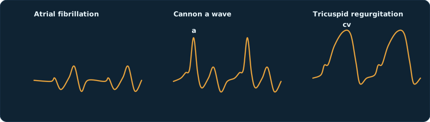
| Pattern | Looks like | Seen in |
|---|---|---|
| Absent a waves | no wave after where the P wave should be | atrial fibrillation |
| Cannon a waves | very tall a waves, regular or intermittent | atrium contracting against a closed tricuspid valve: complete heart block, AV dissociation, ventricular tachycardia, junctional rhythm, ventricular pacing |
| Large a waves | tall a waves every beat | resistance to right atrial emptying: tricuspid stenosis, pulmonary hypertension, a stiff right ventricle |
| Giant cv wave | c and v merge into one large wave and the x descent disappears | tricuspid regurgitation |
| Blunted y descent | slow, shallow fall after the v wave | cardiac tamponade |
| Steep x and y descents | an M or W shape | constrictive pericarditis |
Central line basics
A central venous catheter (CVC, or central line) ends in a large central vein. It is usually placed in the internal jugular (IJ), subclavian, or femoral vein, or threaded from an arm vein as a PICC (peripherally inserted central catheter). Central lines carry vasopressors, irritant drugs, and parenteral nutrition, allow frequent blood draws, and, connected to a transducer, give the CVP trace on this page.
| Stage | What the 2022 SHEA, IDSA, and APIC prevention guidance calls essential |
|---|---|
| Before insertion | a checklist, hand hygiene, and an all-inclusive line kit; in the ICU the subclavian site is preferred to reduce infection |
| Insertion | ultrasound guidance; maximal sterile barriers (cap, mask, sterile gown and gloves, and a full-body drape); skin prep with alcoholic chlorhexidine of at least 2%, allowed to dry |
| Maintenance | chlorhexidine-containing dressings; transparent dressings changed at least every 7 days and gauze every 2 days, sooner if soiled, loose, or damp; hubs disinfected before every access; nonessential lines removed |
Placement is confirmed by a venous, not arterial, waveform when the line is transduced and, for neck and chest lines, by a chest X-ray that shows the tip position and rules out a pneumothorax. The main complications are arterial puncture, pneumothorax, bleeding, air embolism, thrombosis, and central line-associated bloodstream infection (CLABSI).
What CVP cannot tell you
CVP was long used to judge whether a patient needs fluid, but neither a single value nor its change after a fluid challenge reliably predicts whether cardiac output will rise. Read it alongside blood pressure, perfusion, ultrasound, and dynamic measures, and use the waveform for what it does well: rhythm, valve, and pericardial clues, and confirming that a catheter is venous.
How it reads in a note
| As written | Read as |
|---|---|
| R IJ TLC placed under US guidance, tip at cavoatrial junction on CXR | right internal jugular triple-lumen catheter placed with ultrasound; tip position confirmed on chest X-ray |
| CVP 14, large v waves, c/w TR | central venous pressure 14 mmHg with large v waves, consistent with tricuspid regurgitation |
| CVC transduced: venous waveform confirmed | the line was connected to a transducer and shows a venous, not arterial, tracing |
Practice typing it
ICU notes and lines Cardiac ICU
Preset links open the trainer with those settings already applied; you can change them afterward.
See also the arterial line waveform and ventilator settings and waveforms guides.
Common questions
What do the a, c, and v waves mean?
The a wave is right atrial contraction, just after the P wave. The c wave is the tricuspid valve bulging back into the atrium as the ventricle starts to contract, just after the QRS. The v wave is the atrium filling against the closed tricuspid valve, just after the T wave.
What causes cannon a waves?
The right atrium contracting against a closed tricuspid valve. It happens when atria and ventricles lose their normal timing: complete heart block, other forms of AV dissociation, ventricular tachycardia, junctional rhythms, and ventricular pacing.
What does tricuspid regurgitation do to the CVP waveform?
Blood thrown back into the atrium during ventricular systole fills in the x descent and produces a large merged cv wave that can approach right ventricular systolic pressure.
Does CVP predict fluid responsiveness?
Poorly. A single CVP value, or its change after a fluid challenge, does not reliably predict whether cardiac output will rise with more fluid. The waveform is more useful for rhythm, valve, and pericardial clues and for confirming venous placement.
When in the breathing cycle is CVP measured?
At end-expiration, when intrathoracic pressure is closest to its baseline, with the transducer leveled at the phlebostatic axis and zeroed to air.
Sources
- Deranged Physiology. Interpretation of the central venous pressure waveform and abnormal CVP waveform patterns (2023).
- Life in the Fast Lane. CVP measurement (2024).
- StatPearls. Physiology, Jugular Venous Pulsation (2023).
- Buetti N, Marschall J, Drees M, et al. Strategies to prevent central line-associated bloodstream infections in acute-care hospitals: 2022 Update. Infect Control Hosp Epidemiol. 2022;43(5):553-569.
Waveforms on this page are schematic drawings made for it, not patient recordings.